Figures & data
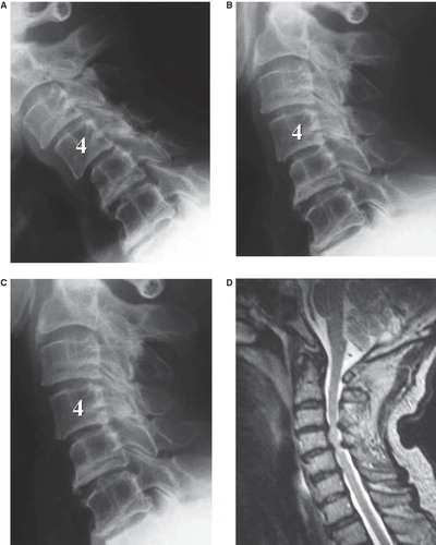
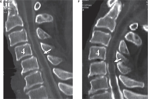
Figure 1. An 84-year-old man with C4/5 myelopathy. A, B, and C: Plain X-ray (A: flexion position, B: neutral position, and C: extension position). In the flexion position, 2-mm anterior slippage in C3 and 3-mm anterior slippage in C4 are demonstrated (A). In the extension position, C3 anterior slippage is reduced, and C4 anterior slippage is 1 mm (C). Space available for the spinal cord at C4/5 in the flexion and extension positions is 12 and 15 mm, respectively. C5-7 range of motion and lordosis angles are 3° and 4°, respectively. D: MRI showed spinal cord compression at C3/4 and C4/5. E and F: CT after myelography (E: flexion position and F: extension position). In the flexion position with maximum anterior slippage and osseous stenosis, deformation due to spinal cord compression is not confirmed (E). In the extension position with minimum anterior slippage and osseous stenosis, findings indicative of spinal cord compression from the posterior direction due to the buckling of the ligamentum flavum are confirmed (F).