Figures & data
Figure 1. Preoperative clinical photography revealed significant scoliosis.
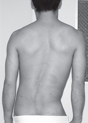
Figure 2. Preoperative plain X-rays. A: Posteroanterior view of the full spine in upright position revealed wedging of the L4/5 disc and the compensatory scoliosis in the cephalad portion of the spine. B: Posteroanterior view of the lumbar spine revealed bilateral spondylolysis of the inferior articular processes of the fourth lumbar vertebra. C: A lateral view of the spine revealed anterolisthesis of the L4 vertebra and a significant pathological opening of the posterior disc space at L4/5 in flexion position.
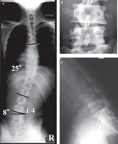
Figure 3. MR images of the lumbar spine revealed spinal canal stenosis at L4/5 (T2-weighted image, sagittal section).
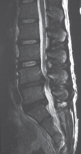
Figure 4. Contrast-enhanced spinal CT scan. A: Frontal plane reconstruction clearly demonstrated spondylolysis of the bilateral inferior articular processes of the L4 vertebra (arrows). B: Horizontal sections at L4/5 disc revealed sagittalization of the L4/5 facet, and deviation of the inferior articular processes of L4 to the right and anterior direction which compressed the dural sac. C: Horizontal sections at the superior margin of the L5 vertebra revealed bone fragments separated from the inferior articular processes of L4.
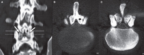
Figure 5. Postoperative clinical photography revealed improvement of scoliosis.
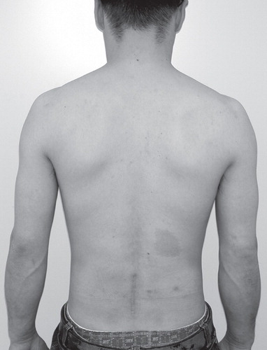
Figure 6. Postoperative plain posteroanterior view of the full spine in upright position revealed bony fusion and improvement of scoliosis.
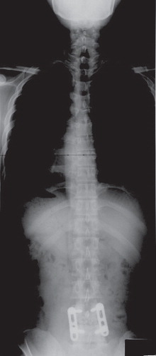
Table I. Reported cases of spondylolysis at articular processes in the lumbar spine.