Figures & data
Figure 1. A lateral radiograph of the right knee. The right patella was enlarged, and its cortical shell was irregularly discontinued. Osteolytic and osteoblastic lesions were irregularly distributed in the patella.
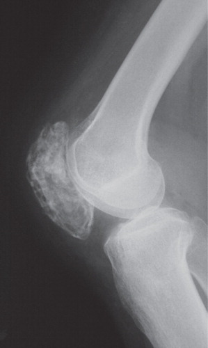
Figure 2. Computed tomography showing an intraosseous osteolytic lesion and soft tissue extension at the anterior part to the patella.
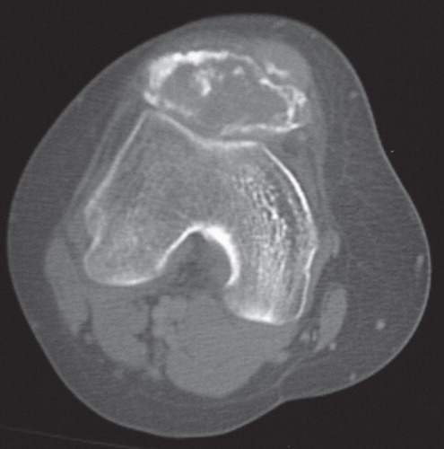
Figure 3. Magnetic resonance imagines showing an intraosseous lesion extending into the anterior soft tissue. A: On T1-weighted images, the lesion showed low signal intensity. B: On T2-weighted images, the proximal part of the lesion showed low signal intensity with partial high-signal areas, and the distal part showed high signal intensity with focal low-signal areas.
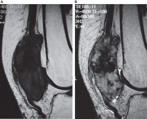
Figure 4. Middle-power view of the biopsied specimens of the patella showing a proliferation of spindle-shaped atypical cells with condensation of chromatin and abundant osteoid formation (×100, hematoxylin and eosin).
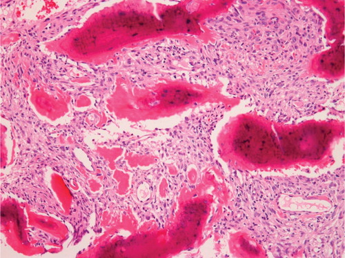
Table I. Details of eight cases on osteosarcoma of the patella.