Figures & data
Figure 1. Well-differentiated neuroendocrine carcinoma of the pancreas (A), with liver metastases (B) (original magnification 400×).
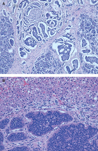
Figure 2. PET scan performed at the end of the fourth cycle of peptide receptor-targeted radionuclide therapy with 177Lu-DOTATATE (A). Gross section of surgical specimens with a circular dark area in the endocervix (yellow arrow) (B).
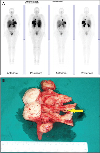
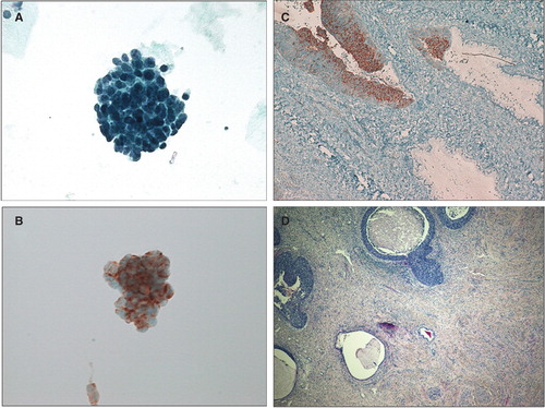
Figure 3. Liquid-based cervical cytology demonstrating atypical cellular clusters (A, B) positive for chromogranin A (B) (original magnification 400×). Histological preparation of the cervix revealing poorly differentiated small cell neuroendocrine tumor involving the cervical crypts (C, D), positive for chromogranin A (C) (original magnification 400×).