Figures & data
Figure 1. Computed tomography of the right thigh of case 1. A circumferential gas pattern was observed.
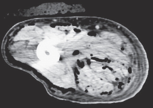
Figure 2. The clinical course and changes in white blood cell count and C-reactive protein of case 1 (survival case). (HBO = hyperbaric oxygen therapy; POD = postoperative day).
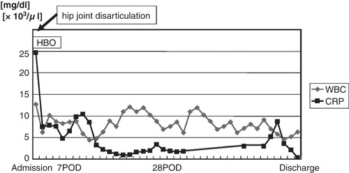
Figure 3. Computed tomography of the left thigh of case 2. A circumferential abscess pattern was observed.
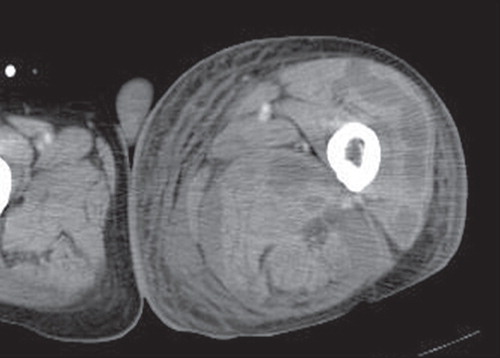
Figure 4. The clinical course and changes in white blood cell count and C-reactive protein of case 2 (death case). (HBO = hyperbaric oxygen therapy; CHDF = continuous hemodiafiltration; POD = postoperative day).
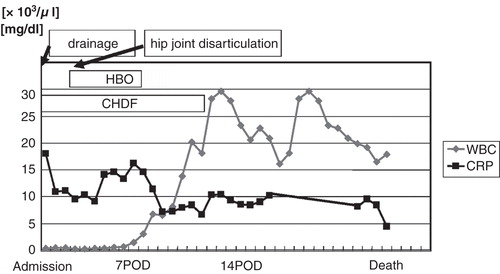
Figure 5. Changes in serum d-ROMs and BAP. Serum d-ROM levels were increased in the patient who survived (case 1, open symbols) but decreased with time in the patient who died (case 2, closed symbols). Serum BAP levels remained within the normal range throughout the course of the disease in both cases. (Adm. = admission). Grey area = normal range
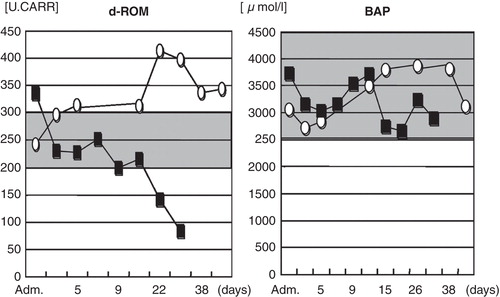
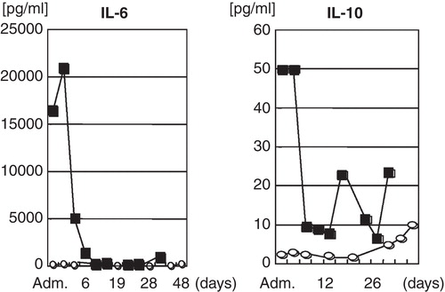
Figure 6. Changes in serum IL-6 (inflammatory cytokine) and IL-10 (anti-inflammatory cytokine). Serum IL-6 and IL-10 were both high on admission in the patient who died (closed symbols); the IL-10 level remained high during hospitalization. (Adm. = admission).