Figures & data
Table I. Total material analyzed for presence of amyloid from patients with lumbar spinal stenosis.
Figure 1. Section of ligament with amyloid deposits immunolabeled for transthyretin and stained with Congo red. Overlapping of immunoreaction with Congo red positivity is evident. Polarized light with partially crossed polars. Bar 200 µm.
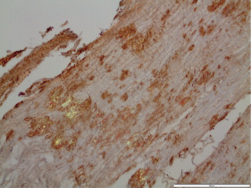
Figure 2. Electron micrograph of ligament with transthyretin-amyloid deposits (A) with typical fine fibrillar appearance, intermingled with collagen fibers (Co). The section was immunolabeled for transthyretin and reaction visualized with 10 nm gold particles. Bar 500 nm.
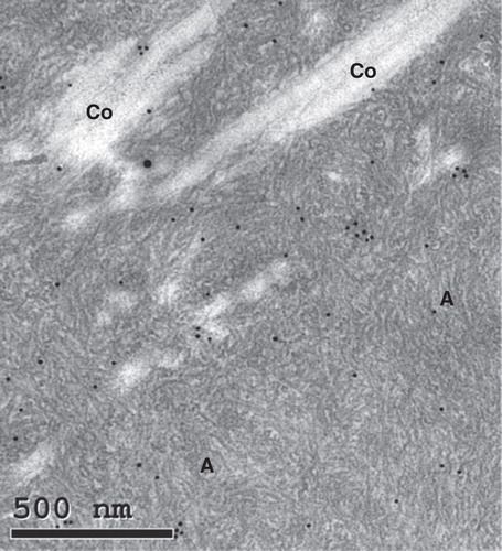
Table II. Amyloid-containing materials with or without reactivity with an anti TTR antibody at immunohistochemistry.
Figure 3. Adjacent sections of ligament immunolabeled for transthyretin (A) and apoA-I (B). While immunolabeling for transthyretin is homogeneous, that for apoA-I is more spotty. Bar 200 µm.
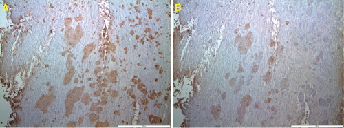
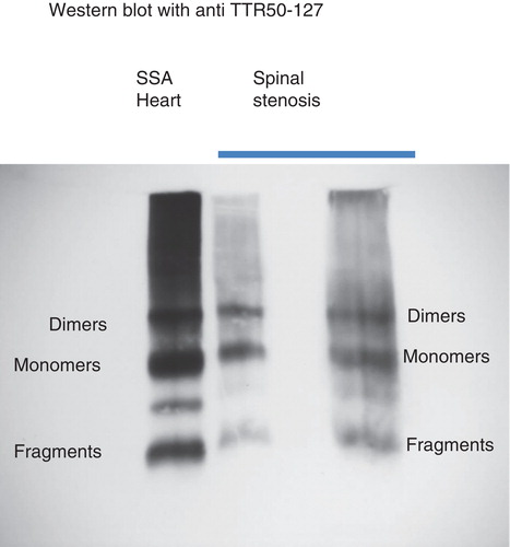
Figure 4. Western blot analysis of extracts of two spinal stenosis materials, positive for transthyretin at immunohistochemistry. Included is also an extract of amyloid fibrils obtained from the heart of a patient with senile systemic amyloidosis. The pattern is the same in all three materials with full-length transthyretin as well as a prominent band corresponding to C-terminal fragments. The unlabeled band between monomers and dimers has constantly been found in senile systemic amyloidosis and may be dimers of fragments.