Figures & data
Figure 1. November 2003. A transverse T2-weighted magnetic resonance imaging (MRI) picture (A) and a coronal T1-weighted one (B), showing a fairly well-circumscribed tumour, measuring about 3 × 3 × 3 cm, located in the soft tissue ventrally to the sacrum, with overgrowth on the peripheral parts of the wall of the rectum. No tumour-like lesions occurred in the sacrum, neither in the coccyx, nor in the cauda equina.
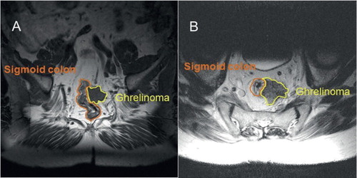
Figure 2. A graphic illustration of the variations in the patient’s blood serum concentrations of total (desacyl-)ghrelin during the time period from June 2009 through August 2013. Those of active (acyl-)ghrelin were consistently non-elevated. The variations were concomitant with those observed in the symptoms and with the clinical effects of the 177Lu treatments.
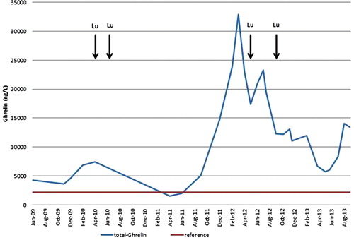
Table I. Survey of the various kinds of treatments received and their effects on the symptoms of the patient’s neoplastic disease.
Figure 3. May 2012. A 68Ga-DOTATATE PET-CT revealed a ‘superscan’ during a progressive phase of the neoplastic disease. There were more than 100 metastatic foci in the skeleton, demonstrating high somatostatin uptake vesicles (SUVs), indicative of somatostatin receptor expression (A: maximum intensity projection image, MIP). Other metastatic sites included liver (B), retro-orbital space (C), kidney (D), pancreas (E), and the femur marrow bilaterally (F). (B–F images in the lower row represent fused PET-CT images corresponding to the CT ones shown in the upper row.)
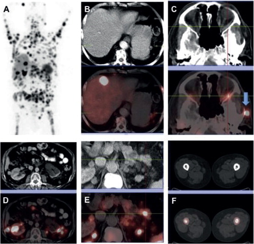
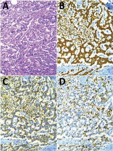
Figure 4. Medium-power (×40) photomicrographs of the neuroendocrine tumour (NET), showing its cellular growth pattern (A) and (in three adjacent sections) its immunoreactivity to antisera raised against pan-cytokeratins (B), CgA (C), and ghrelin (D), respectively.