
Figures & data
Table 1. Characteristics of SLE patients.
Table 2. Characteristics of healthy controls.
Figure 1. Two-group comparisons of (A) EBV EA/D-, (B) CMV pp52-, and (C) HHV6 p41-directed (i) IgM, (ii) IgG, and (iii) IgA levels between SLE patients (n = 77) and healthy controls (n = 29). The antibody levels are presented in arbitrary units (AU). The horizontal bars represent medians and interquartile ranges. *p < 0.05, **p < 0.01, ***p < 0.001.
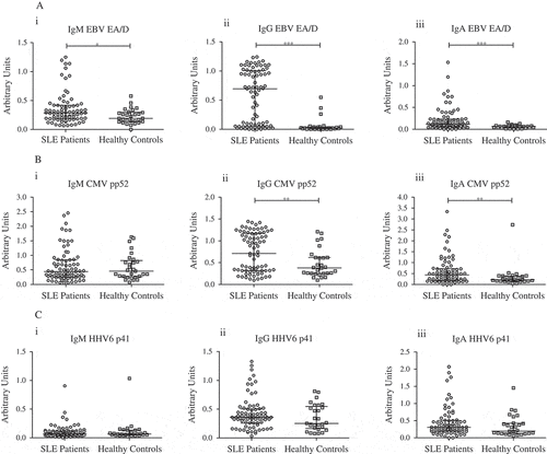
Table 3. Patterns of EBV EA/D- and CMV pp52-directed IgG levels in SLE patients.