Figures & data
Table I. Clinical characteristics of all 58 patients and those with normal (17 patients) or abnormal (25 patients) CSF findings or any MRI pituitary finding (MRI+, 6 patients). Hemoglobin concentration (Hb, g/L), thrombocyte count (×109/L), white blood cell (WBC) count (×109/L), and blood pressure (BP) at the time of hospital entry are shown. The highest values of plasma creatinine (mmol/L) and C-reactive protein (CRP, mg/L) during the hospitalization are given. None of the patients required dialysis.
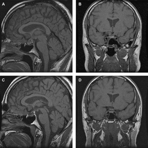
Figure 1. T1-weighted sagittal (A) and coronal (B) magnetic resonance images in a 34-year-old male patient displayed increased signal intensity in the pituitary gland consistent with hemorrhage. The patient developed hypopituitarism in the acute phase of the illness. Three months later (C and D) the pituitary gland had decreased in size, the signal intensity had normalized, as had his hormonal values.