Figures & data
Table I. Definitions for TST positivity, chest imaging classification, and suspected intraocular tuberculosis.
Table II. Epidemiological, clinical and paraclinical investigations for the 108 uveitis patients according to their TST and T-SPOT®.TB status.
Figure 1. Patient with proved tuberculosis. A: Photograph of the fundus showing retinal hemorrhages and occlusive vasculitis of the left eye. B: Fluorescein angiography of the left eye showing vasculitis, with absence of dye filling in some retinal arteries masking retinal hemorrhages. C: Tuberculous cavitation on CT scan. D: Highly positive T-SPOT®.TB test (panel A, ESAT-6 = 177 spots).
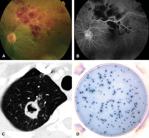
Figure 2. Patient with presumed intraocular tuberculosis and serpiginous choroiditis. A: Fundus photography showing white dots at the choroid, in the posterior pole of the left eye. B: Fluorescein angiography showing hyperfluorescence at the rim of the lesions and hypofluorescence in the center of the lesions. C: Indocyanine angiography showing hypofluorescence of the lesions. D: Highly positive T-SPOT®.TB test (panel B, CFP-10 = 57 spots).
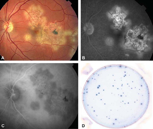
Table III. Epidemiological, clinical, and paraclinical investigations for the 108 uveitis patients included in the study according to their classification as uveitis with determined or undetermined cause, and suspected ocular tuberculosis cases.
Table IV. Test performance of TST, T-SPOT®.TB in the population of 108 uveitis patients (95% confidence interval are noted in brackets).