Figures & data
Figure 1. Mouse adipose organ: anterior subcutaneous depot, interscapular area. Light microscopy of a section where the transition between brown (BAT) and white adipose tissue (WAT) is visible. Note the different cytoplasmic lipid accumulation: mostly unilocular in WAT and multilocular in BAT. Only BAT is intensely stained by UCP1 antibodies (immunohistochemistry ABC method, primary antibody: anti-sheep UCP1 dilution 1:4000, kind gift of Dr D. Ricquier, Paris). Bar = 25 μm.
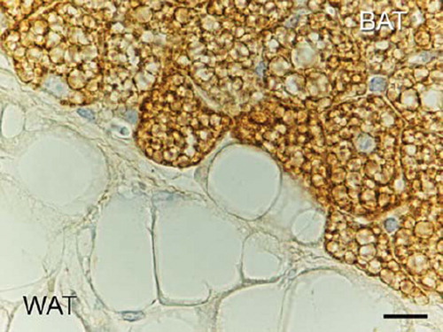
Figure 2. Rat adipose organ: epididymal depot. Electron microscopy of the peripheral part of a white adipocyte. Elongated mitochondria with randomly oriented cristae are visible (arrows). N = nucleus; RER = rough endoplasmic reticulum; large L = main lipid droplet; small L = peripheral lipid droplet; BM = basal membrane. Bar = 0.7 μm.
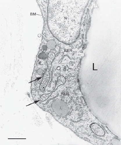
Figure 3. Rat adipose organ: anterior subcutaneous depot, interscapular area. Electron microscopy of cytoplasm of a brown adipocyte. Several big mitochondria packed with cristae are visible. L = lipid droplet (some indicated). Bar = 1 μm.
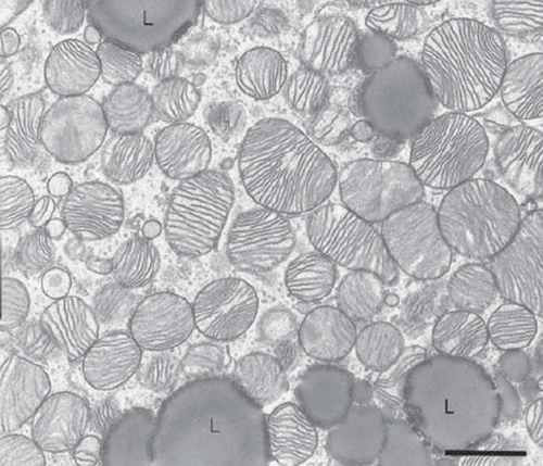
Figure 4. Mouse adipose organ: anterior subcutaneous depot, interscapular area. High-resolution scanning electron microscopy of a cytoplasm of brown adipocyte. 3D aspect of the classic mitochondria. Note the laminar cristae. Bar = 0.4 μm.
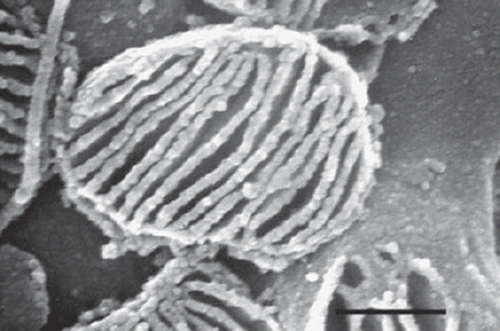
Figure 5. Rat adipose organ: anterior subcutaneous depot, interscapular area. Electron microscopy of a classic neuro-adipose junction. A synapse-like enlargement of a parenchymal nerve fibre is in tight contact with the plasma membrane of a brown adipocyte. V = synaptic vesicles; m = mitochondria (some indicated); N = nucleus of endothelial cell; CAP = capillary lumen. Bar = 0.4 μm.
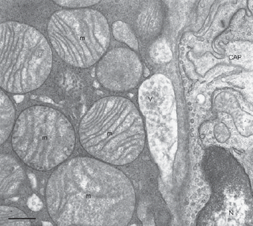
Figure 6. Gross anatomy of mouse adipose organ (adult female Sv129 mice). Middle: mouse kept at warm temperature (28°C for 10 days). Right: mouse kept at cold temperature (6°C for 10 days). Note the different colour of the organ, due to the increase of brown adipose tissue and decrease of white adipose tissue contained in the organ. The organ is made up of two subcutaneous depots (A and F): anterior (A) (composed of: deep cervical, superficial cervical, interscapular, subscapular, axillo-thoracic) and posterior (F) (composed of: dorso-lumbar, inguinal, gluteal); and of several visceral depots: mediastinal (B), mesenteric (C), retroperitoneal (D), and abdomino-pelvic (composed of: perirenal, periovarian, parametrial, perivesical) (E); reproduced from Murano et al., Adipocytes. 2005;1(2):121–30, with permission (Citation36). Bar = 1 cm.
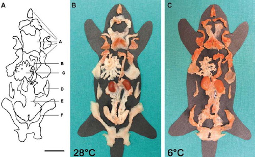
Figure 7. Mouse adipose organ: anterior subcutaneous depot, interscapular area. Immunofluorescence multiple labelling showing parenchymal noradrenergic nerve fibres (tyrosine hydroxylase immunoreactive: red) in tight association with UCP1 immunoreactive brown adipocytes (green) with nuclei in blue (TOTO nuclear counterstaining). Bar = 6 μm.
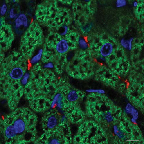
Figure 8. Mouse adipose organ (adult Sv129 female): anterior subcutaneous depot at the periphery of the interscapular area (i.e. white adipose tissue surrounding the classic interscapular brown adipose tissue). UCP1 immunohistochemistry as in . Three days of cold exposure (6°C). Newly formed brown adipocytes appear among white adipocytes. In this depot some newly formed brown adipocytes appear to differentiate from preadipocytes forming small multilocular UCP1 immunoreactive cells (arrows). Some appear to derive from direct transformation of white adipocytes with intermediate stages: UCP1-negative paucilocular cells (asterisk) and UCP1-positive paucilocular cells (double asterisk). Bar = 16 μm.
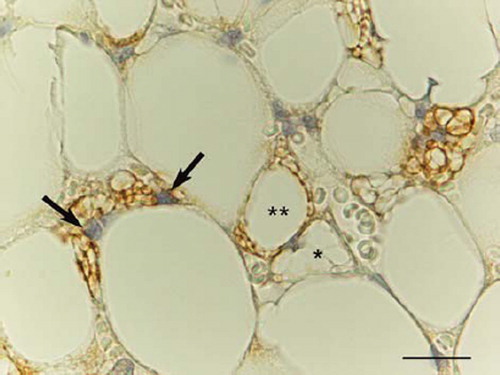
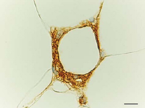
Figure 9. Obese mouse (db/db) adipose organ: omentum. MAC2/Galectin 3 immunoreactive macrophages surround a dead adipocyte forming a classic crown-like structure (CLS). Bar = 15 μm.