Figures & data
Figure 1. Echocardiographic appearance of IVNC. End-diastolic still frame (close-up view of LV apex) of a 37-year-old woman showing the typical echocardiographic features of IVNC with a thin epicardial and a thickened endocardial layer with prominent deep recesses. Blood flow from the ventricular cavity into the recesses is visualized with color Doppler. Reprinted from Steffel et al. (Citation12), Electrocardiographic characteristics at initial diagnosis in patients with isolated left ventricular noncompaction, American Journal of Cardiology 2009;104:984–89, Reprinted from American Journal of Cardiology, volume 104, Steffel J, Kobza R, Oechslin E, Jenni R, Duru F, Electrocardiographic characteristics at initial diagnosis in patients with isolated left ventricular noncompaction, pages 984–89, 2009 (Citation12), Copyright (2009), with permission from Elsevier.
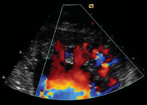
Table I. Electrocardiographic findings in patients with IVNC. Number of patients and (%) are shown.
Table II. Frequency of ventricular arrhythmias in IVNC.
Table III. EP findings in 24 patients with IVNC. Data from Steffel et al., Europace 2009 (Citation10).
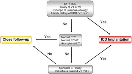
Figure 2. Proposed algorithm for arrhythmic risk stratification in IVNC.
EF = ejection fraction; VT = ventricular tachycardia; VF = ventricular fibrillation; SCD = sudden cardiac death; EDVI = end-diastolic volume index; ICD = implantable cardioverter defibrillator; EP = electrophysiological.