Figures & data
Table I. Baseline characteristics of the study population treated with carvedilol and metoprolol (n=57).
Table II. Effect of 6-month carvedilol and metoprolol treatment on metabolic variables in patients with hypertensive LVH (n=57).
Figure 1. Changes in systolic and diastolic blood pressure and heart rate during treatment with carvedilol and metoprolol for 26 weeks. Comparison of systolic blood pressure (SBP) and diastolic blood pressure (DBP) and heart rate (HR) between baseline and 6-month treatment with cavedilol and metoprolol monitored every 2 weeks.
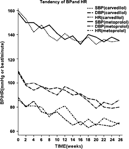
Figure 2. Changes in coronary artery flow velocity in patients with hypertensive left-ventricular hypertrophy during treatment with carvedilol for 26 weeks. At baseline, the peak diastolic and systolic blood flow velocity of the anterior descending coronary artery was 55 and 25 cm/s, respectively (A), and increased to 125 and 60 cm/s, respectively, during maximal hyperemia with intravenous dipyridamole treatment (B). After carvedilol treatment, at baseline the peak diastolic and systolic blood flow velocity was 52 and 20 cm/s, respectively (C), and increased to 160 and 60 cm/s with dipyridamole injection (D). After carvedilol treatment, the ratio of the systolic peak hyperemic flow velocity to baseline flow velocity increased from 2.27 to 2.67 and the ratio of the diastolic peak hyperemic flow velocity to baseline flow velocity increased from 2.4 to 3.0.
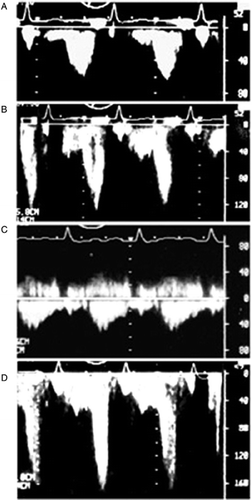
Figure 3. Changes in peak diastolic blood flow velocity (PDV) of anterior descending coronary artery after carvedilol and metoprolol treatment in hypertensive patients with left-ventricular hypertrophy (LVH). PDV at rest and hyperemia increased significantly after carvedilol treatment.
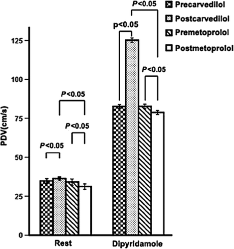
Figure 4. Changes in coronary flow reserve (CFR) before and after 6-month carvedilol and metoprolol therapy in patients with hypertensive left-ventricular hypertrophy (LVH). CFR increased significantly after carvedilol treatment. NS, not significant; M, month.
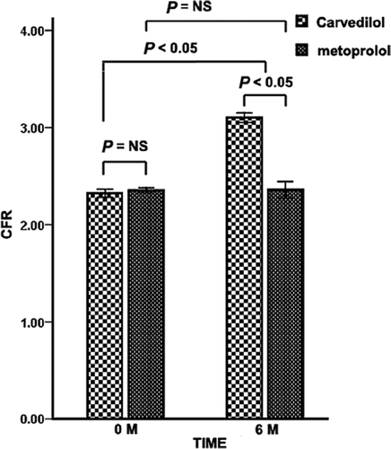
Figure 5. Correlation between change in LV mass index (LVMI) and coronary flow reserve (ΔCFR) after carvedilol treatment in patients with hypertensive LVH. ΔCFR, change in CFR during carvedilol treatment; ΔLVMI, variance in LV mass index during carvedilol treatment. ΔLVMI and ΔCFR have negative correlation after carvedilol treatment.
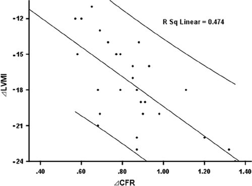
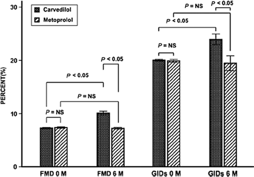
Figure 6. Change in flow-mediated artery dilation (FMD) and glyceryl trinitrate-induced dilation (GID) in brachial artery after 6-month treatment with carvedilol and metoprolol in patients with hypertensive LVH. NS, not significant. M, month.