Figures & data
Figure 1. Influence of an injection of adrenaline into the ICA on the recorded variables (ascending phase): (Citation1) time indicator, (Citation2) amplitude of pial artery pulsation (NIR-T/BSS), (Citation3) blood flow in the left ICA (mL/min), (Citation4) mean BP ( mmHg), (Citation5) respiration, (Citation6) BP in the centripetal portion of the right ICA (mmHg). Initial vasoconstriction, seen as a decrease in pial artery pulsation, which represents the direct response to NE, is followed by passive transmission, seen as an increase in pial artery pulsation, when the BP exceeds the upper autoregulatory limit in the rabbit. Blood flow in the ICA remains diminished. Adapted with permission from Reference 24.
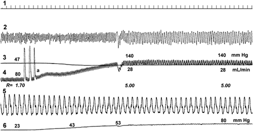
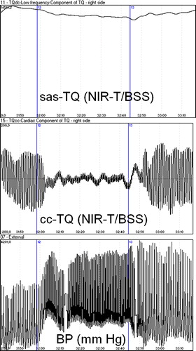
Figure 2. Changes in the width of the subarachnoid space (upper tracing), pial artery pulsation (middle tracing) and mean arterial pressure (lower tracing) during the handgrip test. The start and end of the handgrip test are indicated by vertical lines. Taken with permission from Reference 18.