Figures & data
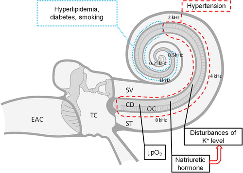
Figure 1. Pure-tone audiometry of a 45-year-old patient with a 7 year history of hypertension: (A) right ear; (B) left ear. Bilateral sensorineural, high-frequency and symmetric hearing loss is present. ○, ×, air conduction; >, <, bone conduction.
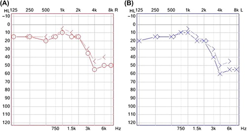
Figure 2. Pathophysiological mechanisms of hypertensive high-tone sensorineural hearing loss. Black dashed line: part of the cochlea affected in arterial hypertension; black dotted line: part of the cochlea additionally affected in the presence of other comorbidities (hyperlipidemia, diabetes) or addictions (smoking). EAC, external auditory canal; TC, tympanic cavity; SV, scala vestibuli; ST, scala tympani; CD, cochlear duct; OC, organ of Corti; K+, potassium ion concentration; pO2, partial pressure of oxygen.