Figures & data
Figure 1. Light microscopic appearance of a renal biopsy demonstrated focal and segmental solidification of glomerular tuft, agglutination of capillaries with hyaline deposits. There is also interstitial infiltrate. H&E × 100.
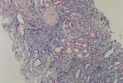
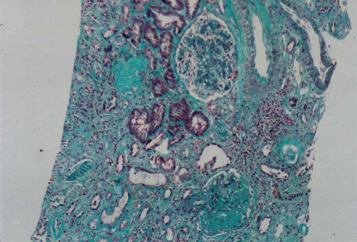
Figure 2. Light microscopic appearance of a renal biopsy showing intense fibrosis. Masson Trichrome staining × 200.