Figures & data
TABLE 1. Semiquantitative comparison of the intensity of TGF-β1 in kidney tissues for each group
TABLE 2. Semiquantitative comparison of the iNOS positive cell numbers in kidney tissues for each group
FIGURE 1. PAS staining of kidney sections of control (a), diabetic untreated (b), diabetic treated with Irb (c), diabetic treated with ALA (d), and diabetic treated with Irb+ALA (e) rats. Increased mesangial matrix, thickened CBMs, TBMs, and GBMs are present in the glomerulus of diabetic untreated rats as compared with the control and diabetic treated rats (arrows: PAS positive area, PAS; scale bar, 50 μm).
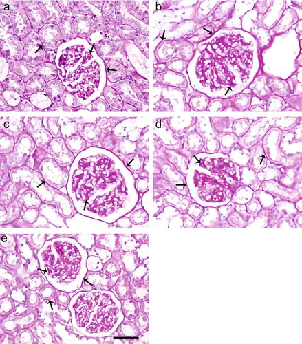
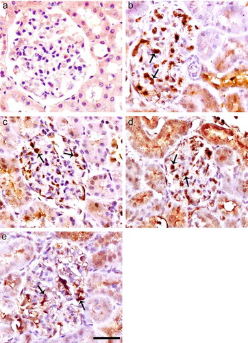
FIGURE 2. Immunoperoxidase staining of TGF-β1 in glomeruli of control (a), diabetic untreated (b), diabetic treated with Irb (c), diabetic treated with ALA (d), and diabetic treated with Irb + ALA (e) rats. Immunostaining of TGF-β1 is increased in glomeruli and tubuli of diabetic rats as compared with the control rats. Immunostaining of TGF-β1 is decreased in glomeruli and tubuli of diabetic-treated rats as compared with the diabetic untreated rats (arrows: positive immunostaining for TGF-β1, immunoperoxidase, hematoxylin counterstain; scale bar, 25 μm).
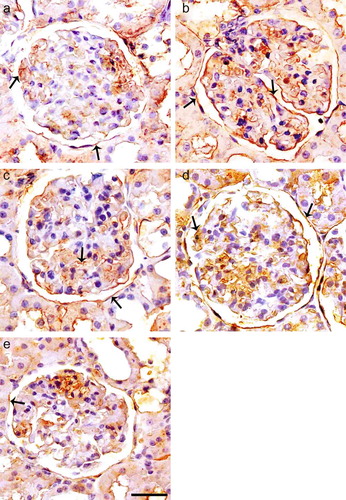
FIGURE 3. Immunoperoxidase staining of iNOS in glomeruli of control (a), diabetic untreated (b), diabetic treated with Irb (c), diabetic treated with ALA (d), and diabetic treated with Irb + ALA (e) rats. Immunohistochemical staining for iNOS is negative in glomerulus of control rats, but positive in the mesangium, podocytes, and capillary loop of untreated diabetic rats. The expression of iNOS in glomerules was lower in ALA and especially Irb-treated diabetic groups as compared with untreated diabetic rats. The expression of iNOS was much lower in ALA treatments in combination with Irb diabetic groups as compared with untreated diabetic group (arrows: positive immunostaining for iNOS, immunoperoxidase, hematoxylin counterstain; scale bar, 25 μm).