781
Views
4
CrossRef citations to date
0
Altmetric
Laboratory Studies

Circulating nucleic acids as possible damage-associated molecular patterns in different stages of renal failure

, , , , , , , , , , , & show all
Pages 486-492 | Received 19 Oct 2009, Accepted 11 Jan 2010, Published online: 06 May 2010

Figures & data

FIGURE 1. (a,b) Spectrophotometric scan analysis of plasma circulating nucleic acids and corresponding standards. For the isolation of RNA from plasma samples, the commercial RNA isolation reagent set TRI Reagent BD (T3809) was used. Purified RNAs and related oligonucleotides were employed for spectrophotometric scan analysis. For spectrophotometric scan analysis the solutions of rRNA, DNA polyA, polyC, polyU, polyI:C, polyA:U, and CpG were used. The UV spectra obtained in our study are in a close agreement with recent revised UV extinction coefficients of RNA and related nucleotides.Citation19

FIGURE 1. (a,b) Spectrophotometric scan analysis of plasma circulating nucleic acids and corresponding standards. For the isolation of RNA from plasma samples, the commercial RNA isolation reagent set TRI Reagent BD (T3809) was used. Purified RNAs and related oligonucleotides were employed for spectrophotometric scan analysis. For spectrophotometric scan analysis the solutions of rRNA, DNA polyA, polyC, polyU, polyI:C, polyA:U, and CpG were used. The UV spectra obtained in our study are in a close agreement with recent revised UV extinction coefficients of RNA and related nucleotides.Citation19

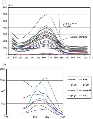
TABLE 1.  Main clinical characteristics of investigated groups and circulating RNA level

FIGURE 2. (a–e) NF-κB, p38, MDA-5, IRF-3, and IRF-7 level after incubation of residential macrophages with isolated nucleic acids. Residential macrophages, cultured in 24-well plates, were allocated into different groups: the cells exposed to media supplemented with physiological saline solution (intact macrophages); the cells exposed to media supplemented with RNAs purified from plasma of control healthy subjects; the cells exposed to media supplemented with RNAs purified from different groups of patients. The number of samples of each group corresponded to the number of circulating nucleic acid samples, except that untreated group of cells exposed to media and physiological saline solution consisted of 10 samples. The residential macrophages were cultured with isolated nucleic acids for 4 hours, afterwards the cells were transferred into five 96-well plates for determination of NF-κB, p38, MDA-5, IRF-3, and IRF-7. The MFI (logarithmic scale) was determined, and the results presented here are the values of MFI following subtraction of values for unstained matched control cells, analyzed on Victor Perkin Elmer–Wallac multiplate reader. *p < 0.001, statistical significance compared to the cells exposed to media supplemented with RNAs purified from plasma of control healthy subjects. **p < 0.001, statistical significance compared to the cells exposed to media supplemented with RNAs purified from plasma of patients who underwent renal transplantation. ***p < 0.05, statistical significance compared to the cells exposed to media supplemented with physiological saline solution (intact macrophages).

FIGURE 2. (a–e) NF-κB, p38, MDA-5, IRF-3, and IRF-7 level after incubation of residential macrophages with isolated nucleic acids. Residential macrophages, cultured in 24-well plates, were allocated into different groups: the cells exposed to media supplemented with physiological saline solution (intact macrophages); the cells exposed to media supplemented with RNAs purified from plasma of control healthy subjects; the cells exposed to media supplemented with RNAs purified from different groups of patients. The number of samples of each group corresponded to the number of circulating nucleic acid samples, except that untreated group of cells exposed to media and physiological saline solution consisted of 10 samples. The residential macrophages were cultured with isolated nucleic acids for 4 hours, afterwards the cells were transferred into five 96-well plates for determination of NF-κB, p38, MDA-5, IRF-3, and IRF-7. The MFI (logarithmic scale) was determined, and the results presented here are the values of MFI following subtraction of values for unstained matched control cells, analyzed on Victor Perkin Elmer–Wallac multiplate reader. *p < 0.001, statistical significance compared to the cells exposed to media supplemented with RNAs purified from plasma of control healthy subjects. **p < 0.001, statistical significance compared to the cells exposed to media supplemented with RNAs purified from plasma of patients who underwent renal transplantation. ***p < 0.05, statistical significance compared to the cells exposed to media supplemented with physiological saline solution (intact macrophages).

Reprints and Corporate Permissions

Please note: Selecting permissions does not provide access to the full text of the article, please see our help page How do I view content?

To request a reprint or corporate permissions for this article, please click on the relevant link below:

Academic Permissions

Please note: Selecting permissions does not provide access to the full text of the article, please see our help page How do I view content?

Obtain permissions instantly via Rightslink by clicking on the button below:

If you are unable to obtain permissions via Rightslink, please complete and submit this Permissions form. For more information, please visit our Permissions help page.