Figures & data
FIGURE 1. Kaplan–Meier curve showing time from initiation of PD to EPS (SP) diagnosis.
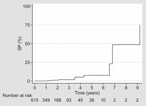
TABLE 1. Multifactorial model to determine risk of EPS
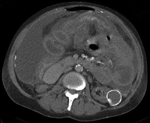
FIGURE 2. CT abdomen demonstrates nodular peritoneal thickening and peritoneal calcification. There are free fluid and loculated fluid collections on the left. The bowel loops are centrally located.