Figures & data
FIGURE 1. A CT scan of the abdomen in a patient on peritoneal dialysis who presented with peritonitis. (A) The first CT scan showed ascites in the abdominopelvic cavity, with some particular peritoneal thickening with enhancement in the pelvic cavity, suggesting possibility of peritonitis. (B) The second CT scan showed increased ascites, peritoneal thickening with enhancement, and thickening of the diffuse bowel loop, suggesting peritonitis and reactive bowel wall thickening, and dilatation and increased enhancement of appendiceal loop (arrow). (C, D) The third CT scan demonstrated perforated appendicitis with peri-appendiceal abscess (arrow).
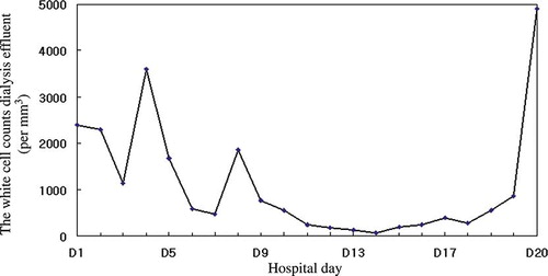
FIGURE 2. The change of white blood cell counts in the dialysis effluent during IP antibiotic treatment.