Figures & data
Figure 1. Glomerulus showing mesangial proliferation with cellular crescent formation (periodic acid–Schiff stain, ×400).
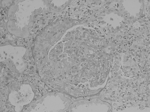
Figure 2. Immunofluorescence staining for IgA showing 2+ granular deposition in mesangium and glomerular capillary loops (original magnification ×400); there is also 2+ staining for C3 in a similar pattern (not shown).
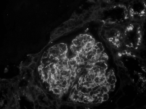
Figure 3. Electron microscopy showing electron-dense deposits in the mesangium (original magnification ×8000).
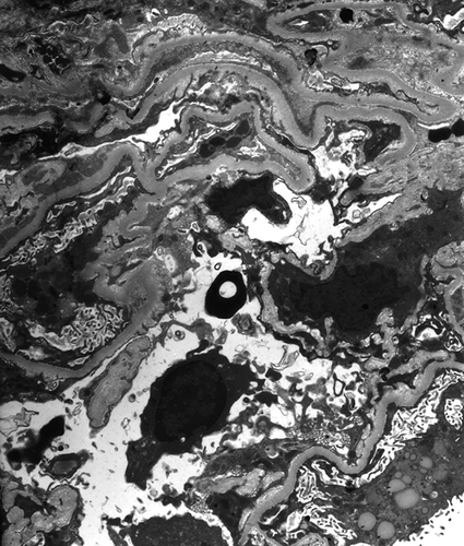
Table 1. Clinical characteristics of patients with crescentic IgA nephropathy associated with staphylococcal infection described in the literature