Figures & data
Table 1. Incidence and types of glomerulonephropathy associated with lymphoma.Citation20
Figure 1. Glomerular basement membrane thickening (yellow arrows) and mononuclear cell infiltration (arrows) in kidney interstitium (HE, ×200). HE, Hematoxylin eosinophil.
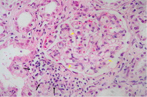
Figure 3. B-cell lymphocytes stained by antibodies CD20 infiltration (arrows) in kidney interstitium (ABP, CD20, ×200). ABP, avidin biotin peroxidase method.
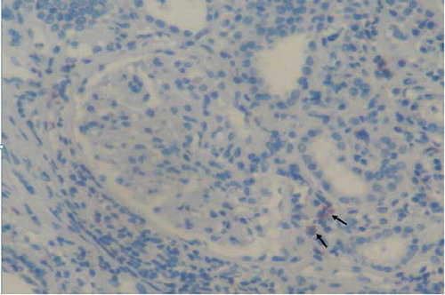
Figure 2. Small T-cell lymphocytes stained by antibodies CD3 infiltration (arrows) in kidney interstitium (ABP, CD3, ×200). ABP, avidin biotin peroxidase method.
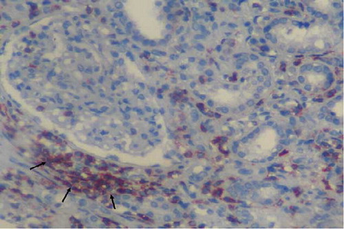
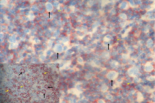
Figure 4. Reed–Sternberg cells and reactive small lymphocytes (arrows) in lymph node sections (HE, ×200). Inset: cytoplasmic CD30 positivity in Reed–Sternberg cells (ABP, CD30, ×400). ABP, avidin biotin peroxidase method. Hematoxylin eosinophil.
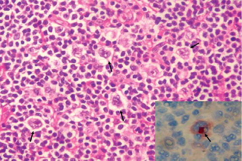
Figure 5. Reed–Sternberg cells (white arrows) and CD3-positive reactive small lymphocytes (yellow arrows) in lymph node sections (ABP, CD3, ×200). Inset: CD20-positive Reed–Sternberg cells (arrows) and CD20-positive reactive small B lymphocytes (yellow arrows) in lymph node sections (ABP, CD20, ×200). ABP, avidin biotin peroxidase method.