Figures & data
Table 1. Physicochemical properties for iodixanol, iohexol, iopromide, and ioversol
Table 2. Cell death (%, mean ± SEM), measured by the trypan blue exclusion assay, in NRK 52-E cells after exposure to incubation medium (control), iodixanol, iohexol, iopromide, and ioversol (150 mg I/mL)
Table 3. Cell viability (percent of control, mean ± SEM), as determined by mitochondrial dehydrogenase activity, in NRK 52-E cells after exposure to incubation medium (control), iodixanol, iohexol, iopromide, and ioversol (150 mg I/mL)
Table 4. Cell death (%, mean ± SEM) assessed on formalin-fixed NRK 52-E cells after exposure to incubation medium (control), iodixanol, iohexol, iopromide, and ioversol (150 mg I/mL) (n = 5: two counts from the first specimen and three from the last)
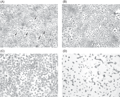
Figure 1. (A) Hematoxylin-stained NRK 52-E cells (control – incubation media treated) (×200). Note the confluence of the cell layer. Randomly scattered throughout the culture are cells that have separated from the surrounding cells and contain condense dark nuclei (arrows). These are degenerating/dying cells. (B) Hematoxylin-stained NRK 52-E cells after 1 h exposure to 150 mg I/mL ioversol (×400). Note the increased number of dead cells scattered throughout the cell culture layer. (C) Hematoxylin-stained NRK 52-E cells after 3 h exposure to 150 mg I/mL ioversol (×400). There are more necrotic cells and the confluence of the cell culture layer is lost. (D) Hematoxylin-stained NRK 52-E cells after 12 h exposure to 150 mg I/mL ioversol (×400). Note that almost all the cells are either necrotic or dying. Also cell-to-cell contact is minimal.