Figures & data
Figure 1. Eosinophilic hyaline deposits were seen in the mesangium and afferent arteriolar wall. Jones methenamine silver stain, original magnification ×400.
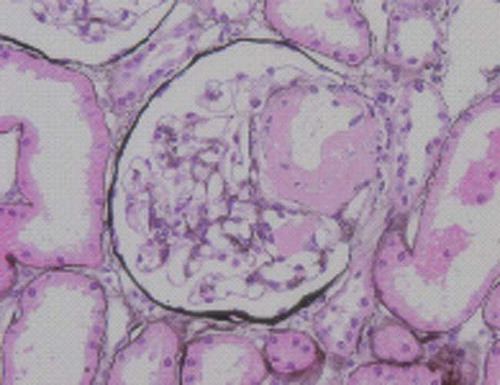
Figure 2. Electron micrograph showing fibrillary deposits in the mesangium. Deposits were formed by randomly arranged fibrils. Original magnification ×100,000.
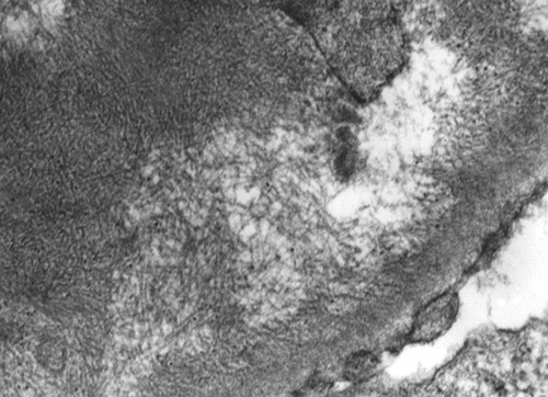
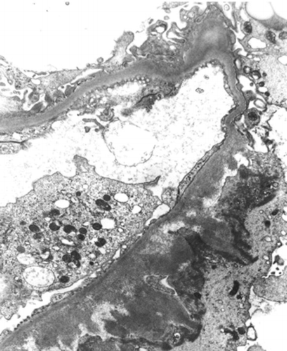
Figure 3. Electron micrograph showing fibrillary deposits in the sub-epithelial zone of the capillary wall. Original magnification ×5000.