Figures & data
Figure 1. Light microscopy findings. The glomerulus exhibits segmental sclerosis without vacuolated cells (PAS staining, original magnification ×300).
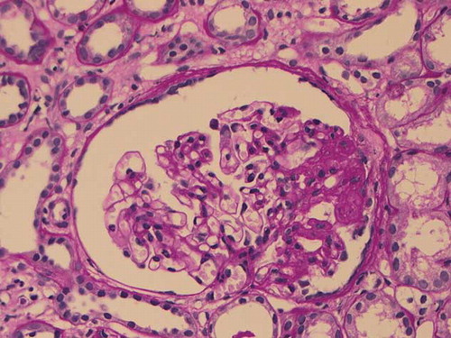
Figure 2. Electron microscopy findings. Myeloid bodies are seen in the glomerular epithelial cells. A few mesangial inclusions are also present (original magnification ×2150).
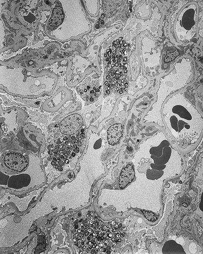
Table 1. The characteristics of patients with glomerular myeloid bodies without Fabry’s disease.