Figures & data
Figure 1. Magnetic resonance urography revealed calyceal dilatation, increased number of calyces, and normal ureters and bladder.
Figure 2. Glomeruli had no significant alterations light microscopically [(A) hematoxylin and eosin stain, ×200 magnification and (B) Masson’s trichrome stain, ×400 magnification].
![Figure 2. Glomeruli had no significant alterations light microscopically [(A) hematoxylin and eosin stain, ×200 magnification and (B) Masson’s trichrome stain, ×400 magnification].](/cms/asset/5e57354d-ed17-49e7-aa8b-ddb5c315cdf0/irnf_a_731996_f0002_b.jpg)
Figure 3. Mild tubular atrophy with interstitial fibrosis is noted at right side of the picture (Jone’s methanamine silver stain, × 200 magnification). Inset shows deposition of IgA in the mesangial regions (imunofluorescence, fluorescein isothiocyanate-conjugated anti-IgA antibody ×400).
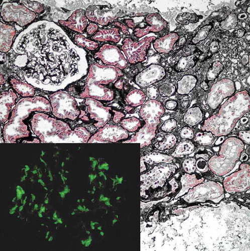
Table 1. Clinical and demographic characteristics of the cases with congenital megacalycosis described in the literature.