Figures & data
Figure 1. Hematoxylin and eosin staining of the radial artery.
Notes: The calcification was located in the tunica media. The calcificated endangium was basically complete, and the thickening of tunica media can be seen and was significantly correlated with the increase of calcification. The differences among noncalcification, mild to moderate calcification, and severe calcification groups were statistically significant (p < 0.05).
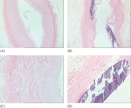
Table 1. Thickness of tunica media in radial artery and IOD values for immunohistochemical staining (n = 80, x ± s).
Figure 2. Alizarin red staining of radial artery.Note: The purple or orange-red calcium deposits can be seen in the calcificated tunica media.
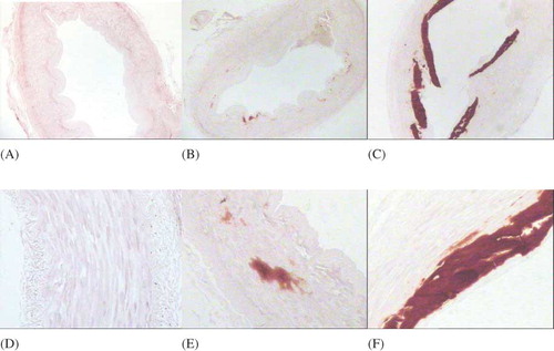
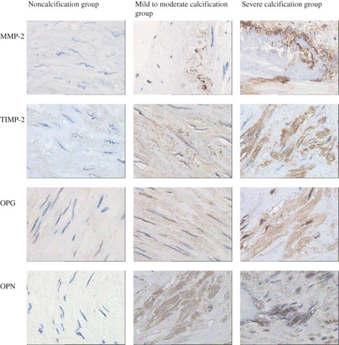
Figure 3. Immunohistochemical staining of radial artery.Notes: The expression of MMP-2, TIMP-2, OPG, and OPN can be seen in the ×400 calcificated radial artery. The expression increased with the deepening of the degree of vascular calcification, with statistically significance (p < 0.05).