950
Views
17
CrossRef citations to date
0
Altmetric
Case Report

Acute Kidney Injury by Arsine Poisoning: The Ultrastructural Pathology of the Kidney

, , , , &
Pages 299-301 | Received 09 Oct 2012, Accepted 23 Oct 2012, Published online: 30 Nov 2012

Figures & data

Figure 1.  The change of serum creatinine levels during hospitalization.

Figure 1.  The change of serum creatinine levels during hospitalization.

Figure 2.  Light microscopy shows that the glomerulus appears normal and tubular epithelial cells are degenerated with focal loss of nuclei. Some tubular lumens are dilated and filled with eosinophilic casts. Interstitia are slightly widened with a few mononuclear cells (H&E, ×200).
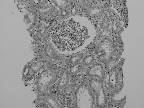
Figure 2.  Light microscopy shows that the glomerulus appears normal and tubular epithelial cells are degenerated with focal loss of nuclei. Some tubular lumens are dilated and filled with eosinophilic casts. Interstitia are slightly widened with a few mononuclear cells (H&E, ×200).

Figure 3.  The section shows many lysosomes containing dense granular materials (black arrow). Equal-sized oval mitochondria are considered to be normal (white arrow). Diverse-shaped and small-sized mitochondria that have a dense matrix are considered to be degenerated (hollow arrow) (second male, electron microscopy, ×10,000).
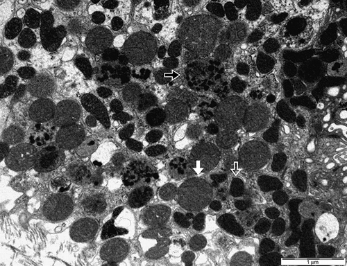
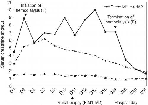
Figure 3.  The section shows many lysosomes containing dense granular materials (black arrow). Equal-sized oval mitochondria are considered to be normal (white arrow). Diverse-shaped and small-sized mitochondria that have a dense matrix are considered to be degenerated (hollow arrow) (second male, electron microscopy, ×10,000).
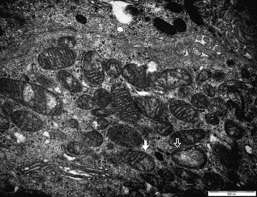
Figure 4.  The section shows that the outer membranes of mitochondria are ruptured and the cristae are disrupted (white arrow). A few mitochondria show vacuolated changes (hollow arrow) (first male, electron microscopy, ×20,000).

Figure 4.  The section shows that the outer membranes of mitochondria are ruptured and the cristae are disrupted (white arrow). A few mitochondria show vacuolated changes (hollow arrow) (first male, electron microscopy, ×20,000).

Reprints and Corporate Permissions

Please note: Selecting permissions does not provide access to the full text of the article, please see our help page How do I view content?

To request a reprint or corporate permissions for this article, please click on the relevant link below:

Academic Permissions

Please note: Selecting permissions does not provide access to the full text of the article, please see our help page How do I view content?

Obtain permissions instantly via Rightslink by clicking on the button below:

If you are unable to obtain permissions via Rightslink, please complete and submit this Permissions form. For more information, please visit our Permissions help page.