Figures & data
Table 1. Methods used to assess kidney function.
Table 2. Patient characteristics.
Table 3. Measured GFR and surrogate estimates of kidney function by patient (expressed in mL/min/1.73 m2).
Figure 1. Box plot of the difference in the mean 125I-Urine and other assessment methods. The line within the box marks the median. Error bars above and below the box indicate the 90th and 10th percentiles.
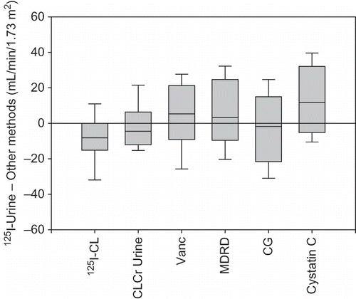
Figure 2. Bland–Altman plots of the relationship between 125I-Urine and (A) 125I-CL; (B) CLCr Urine; (C) V-CL; (D) MDRD; (E) CG-TBW; and (F) Cystatin C. Solid bar indicates the mean difference or bias. Dashed lines indicate the upper and lower limits of the interval of agreement (±1SD).
