Figures & data
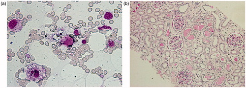
Figure 1. Clinical course of the patient. Notes: She developed complete anuria on hospital day 4. The multiple combination therapy, including steroid, cyclosporine, etoposide, and plasmapheresis, failed. She was always febrile over 37 °C and even just before her death, the level of serum IL-6 and soluble IL-2R were very high. Abbreviations: PSL = prednisolone, m-PSL = methylprednisolone sodium-succinate, HD = hemodialysis, HDF = hemodiafiltration, PE = plasma exchange, BT = body temperature, SAH = subarachnoid hemorrhage, n.d. = not data.
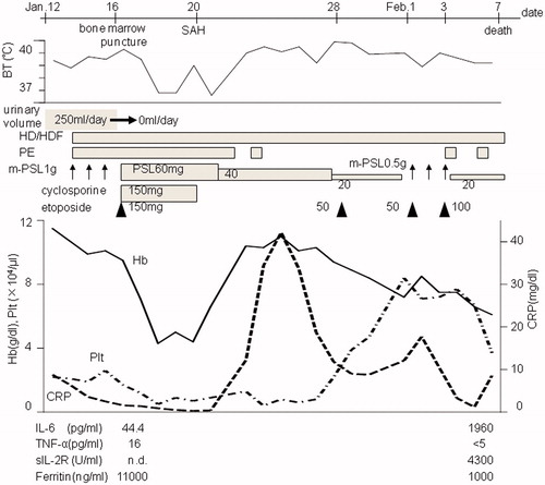
Figure 2. (a) Wright–Giemsa staining of the bone marrow aspirate. There are two macrophage-phagocytosing erythrocytes and platelets (original magnification ×400). (b) Periodic acid-Schiff staining of renal necropsy specimen. There is some cell infiltration in the interstitium and no acute tubular necrosis, although detachment of tubular cells due to postmortem changes was detected. There is remarkable edema in the interstitium and some protein casts with intact glomeruli (original magnification ×100).