Figures & data
Table 1. Clinical and histopathologic data (n = 35).
Figure 1. (a) and (b): Immunohistochemical staining of basal cells with SP-A and SP-D proteins (original magnification was 200×).
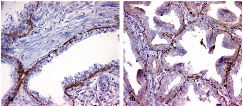
Figure 2. Strong positive SP-A (a) and SP-D (b) immunostaining particularly in inflammatory sites of non-malignant prostate tissue specimens. Note: Original magnifications for the images was 200×.
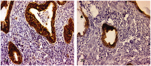
Figure 3. Immunohistochemical staining intensity scores. Prostate adenocarcinomas and non-malignant areas in same tissue sections stained with anti SP-A antibody (a) 1+ staining, (c) 2+ staining (e) 3+ staining. Non-malignant areas stained with anti SP-A antibody (b) 1+ staining, (d) 2+ staining (f) 3+ staining. Negative staining (nc1) of prostate adenocarcinoma, negative control staining of non-malignant areas (nc2). Note: Original magnifications of images; (a) 200×, (b) 50×, (c) 100×, (d) 100×, (e) 50×, (f) 50×, nc1; 50×, nc2: 100×.
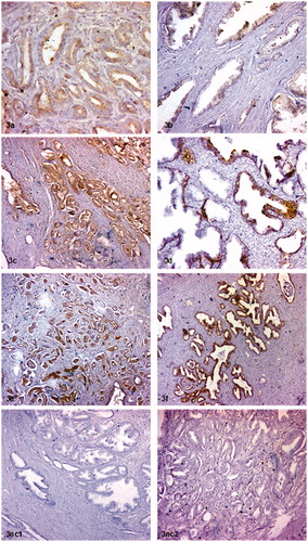
Figure 4. Immunohistochemical staining intensity scores. Prostate adenocarcinomas and non-malignant areas in same tissue sections stained with anti SP-D antibody (a) 1+ staining, (c) 2+ staining. Non-malignant areas stained with anti SP-D antibody (b) 1+ staining, (d) 2+ staining (e) 3+ staining. Negative control staining (nc). Note: Original magnifications of images; (a) 100×, (b) 50×, (c) 50×, (d) 50×, (e) 100, nc 50×.
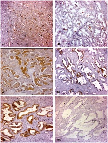
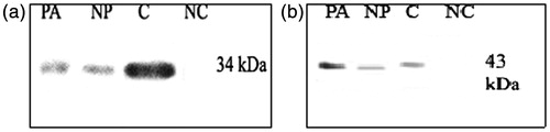
Figure 5. (a) and (b) Western blot analysis of SP-A and SP-D in prostate from control and prostate adenocarcinoma. A. Western blot of prostate extracts obtained from noncadenocarcinomas site (NP) and adenocarcinoma field (PA) were probed with antibodies against SP-A (a) and SP-D (b). Note: PA; prostate adenocarcinoma, NP; normal prostate, C; control, NC; negative control.