Figures & data
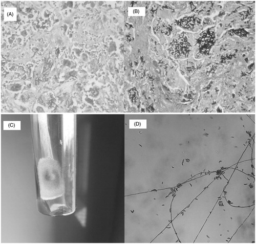
Figure 1. (A) Histopathology- granuloma with giant cell studded with fungal elements (H & E stain). (B) Histopathology- intracellular fungal hyphae (Gomori’s methenamine silver stain). (C) Growth on Sabouraud agar – fungal colony with cottony surface. (D) Growth on Sabouraud agar – banana-shaped macroconidia.