
Figures & data
Table 1. Clinical characteristic at diagnosis of six children with distal renal tubular acidosis.
Table 2. Hearing status and inner ear radiologic characteristics of these children with distal renal tubular acidosis.
Figure 1. Novel mutations identified in ATP6V1B1 and ATP6V0A4 of patients with distal renal tubular acidosis. The mutant nucleotides are marked with asterisks.
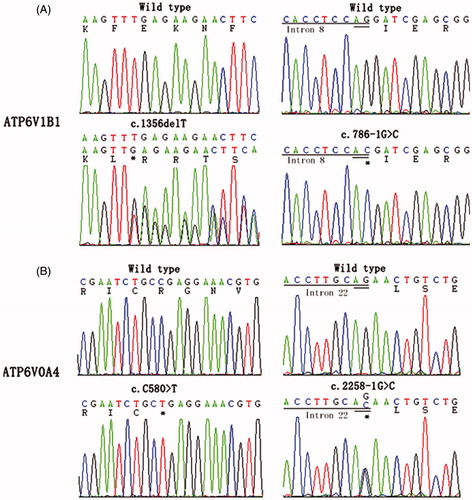
Table 3. Detected mutations in ATP6V1B1 and ATP6V0A4 of the six children with distal renal tubular acidosis.
Figure 2. High-resolution computed tomography (CT) and magnetic resonance imaging (MRI) of the temporal bones in patient III-2. Bilateral enlargement of the vestibular aqueduct and endolymphatic sac (arrows) is evident.
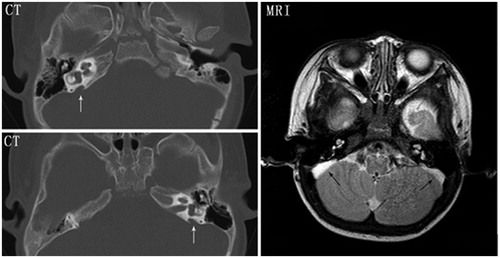
Figure 3. Audiograms of patients III-2 and IV-2. (A) Audiogram of patient III-2 at age of 5 years. (B) Audiogram of patient III-2 at age of 8 years. (C) Audiogram of patient IV-2 at age 5 years. Bilateral air–bone gaps are recognized, especially at lower frequencies in patient III-2. Circle or × indicate unmasked examination of air conduction for the right or left ear, triangle or square indicate masked examination of air conduction for right or left ear, < or > indicate unmasked bone conduction for right or left ear, [ or ] indicate masked bone conduction for right or left ear.
![Figure 3. Audiograms of patients III-2 and IV-2. (A) Audiogram of patient III-2 at age of 5 years. (B) Audiogram of patient III-2 at age of 8 years. (C) Audiogram of patient IV-2 at age 5 years. Bilateral air–bone gaps are recognized, especially at lower frequencies in patient III-2. Circle or × indicate unmasked examination of air conduction for the right or left ear, triangle or square indicate masked examination of air conduction for right or left ear, < or > indicate unmasked bone conduction for right or left ear, [ or ] indicate masked bone conduction for right or left ear.](/cms/asset/a956e013-ecf2-4d56-9567-52e4f799c828/irnf_a_930332_f0003_b.jpg)