Figures & data
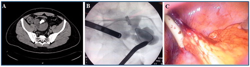
Figure 1. Approximately 30 mm calculus in the renal pelvis of malrotated right kidney (A); renal access was performed directly into the renal pelvis under laparoscopic and fluoroscopic control (B); complete stone clearence achieved and a double-J stent was inserted (C) 301 × 94mm (96 × 96 DPI).
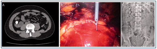
Figure 2. Partial staghorn renal calculi in right pelvic ectopic kidney (A); under laparoscopic vision and fluoroscopic control, percutaneous access to the lower calix was obtained (B); a nephrostomy tube was inserted (C). 266 × 70mm (96 × 96 DPI).