Figures & data
Figure 1. The histological scores of all the groups. Values are mean ± SD for six rats in each group. (a,b,c): Values with common superscripts are not statistically different, whereas values without common superscripts are statistically significantly different (p value <0.05).
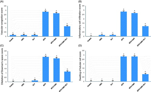
Figure 2. Photomicrographs of kidney sections stained with hematoxylin and eosin (scale bars = 20 μm), showing: (A) Group 1 (control), (B) Group 2 (CMC) and (C) Group 3 (SLY) show similarly undamaged kidney; (D) Group 4 (MTX) dilatation of Bowman’s space (arrow) and inflammatory cell infiltration (asterisk); (E) Group 4 (MTX) glomerular (arrows) and peritubular (asterisks) vascular congestion; (F) Group 4 (MTX) swelling of renal tubular epithelium cells (arrows); (G) Group 5 (MTX + CMC) inflammatory cell infiltration (asterisk); (H) Group 5 (MTX + CMC) dilatation of Bowman’s space (thick arrow) and glomerular (thin arrow), peritubular (asterisks) vascular congestion; (I) Group 5 (MTX + CMC) swelling of renal tubular epithelium cells (arrows); (J) Group 6 (MTX + CMC + SLY) inflammatory cell infiltration (asterisk); (K) Group 6 (MTX + CMC + SLY) glomerular (thin arrow) and peritubular (asterisk) vascular congestion, Bowman’s space (thick arrow); (L) Group 6 (MTX + CMC + SLY) swelling of renal tubular epithelium cells (arrows).
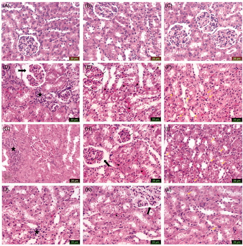
Figure 3. The apoptotic index of all the groups. Values are mean ± SD for six rats in each group. (a,b,c): Bars with common superscripts are not statistically different, whereas values without common superscripts are statistically significantly different (p value <0.05).
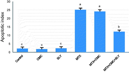
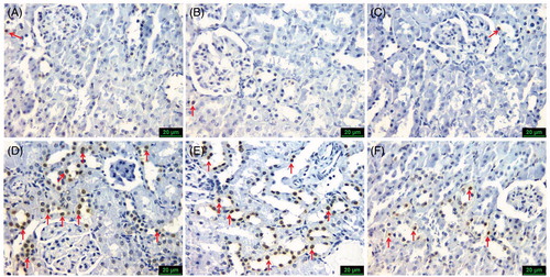
Figure 4. Representative photomicrographs of TUNEL staining in all six groups (scale bars=20 μm), showing: (A) Group 1 (control), (B) Group 2 (CMC) and (C) Group 3 (SLY) similarly only few TUNEL-positive cells (arrow); (D) Group 4 (MTX) and (E) Group 5 (MTX + CMC) similarly a lot of TUNEL-positive cells (arrows); (F) Group 6 (MTX + CMC + SLY) rare TUNEL-positive cells (arrows).