973
Views
3
CrossRef citations to date
0
Altmetric
Clinical Study

Ultrasonographic evaluation of the femoral cartilage thickness in patients with chronic renal failure

, , , , , & show all
Pages 600-604 | Received 08 Dec 2015, Accepted 26 Jan 2016, Published online: 24 Feb 2016

Figures & data

Figure 1. Ultrasonographic images (suprapatellar axial view) showing bilateral femoral cartilage measurements. ICA: intercondylar area; L: left; LFC: lateral femoral condyle; MFC: medial femoral condyle; R: right.

Figure 1. Ultrasonographic images (suprapatellar axial view) showing bilateral femoral cartilage measurements. ICA: intercondylar area; L: left; LFC: lateral femoral condyle; MFC: medial femoral condyle; R: right.

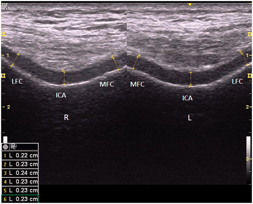
Table 1. The demographic and clinical characteristics of the CRF patients and controls.

Table 2. Comparison of the femoral cartilage thickness measurements (mm) of the CRF patients and the healthy controls (mean ± standard deviation).

Reprints and Corporate Permissions

Please note: Selecting permissions does not provide access to the full text of the article, please see our help page How do I view content?

To request a reprint or corporate permissions for this article, please click on the relevant link below:

Academic Permissions

Please note: Selecting permissions does not provide access to the full text of the article, please see our help page How do I view content?

Obtain permissions instantly via Rightslink by clicking on the button below:

If you are unable to obtain permissions via Rightslink, please complete and submit this Permissions form. For more information, please visit our Permissions help page.