
Figures & data
Figure 1. (A) H&E stain (×200) showing increased mesangial hypercellularity in a glomerulus. (B) Silver stain (×400) showing segmental proliferative lesions in a glomerulus.
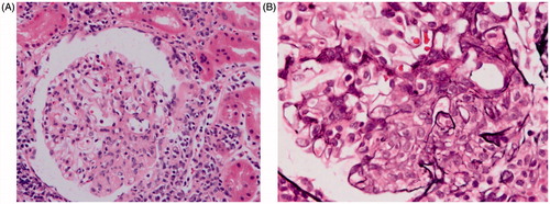
Figure 2. Electron microscopy of the kidney biopsy showing mesangial immune dense deposits.
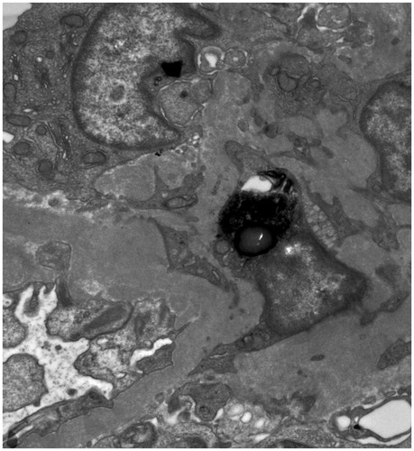
Figure 3. CT image of the left kidney showing perirenal hematoma.
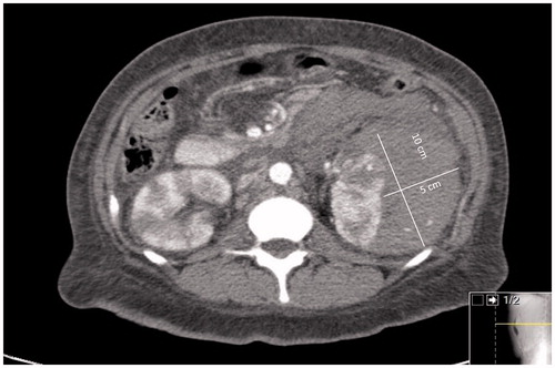
Figure 4. (A) Angiogram of the left kidney showing multiple microansurysms. (B) Coil placement in a renal artery branch feeding into the hemoatoma [white arrow].
![Figure 4. (A) Angiogram of the left kidney showing multiple microansurysms. (B) Coil placement in a renal artery branch feeding into the hemoatoma [white arrow].](/cms/asset/3c3feaa6-93fe-4809-9dab-4bf464bbe99d/irnf_a_1165075_f0004_c.jpg)