Figures & data
FIGURE 1 Fundus photographs (A) and fluorescein angiogram (FA) (B). Retinal pigment epithelial changes and foveal granularity are seen in the right eye (left), and the left fundus (right) is normal (A). FA of the right eye reveals pinpoint hyperfluorescent spots that stain late (B).
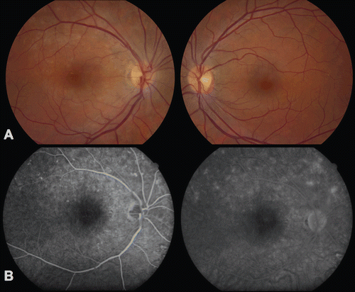
FIGURE 2 Spectral Domain Optical Coherence Tomography ((SD-OCT) Spectralis; Heidelberg Engineering, Germany), right eye. Baseline OCT shows disruption of the inner segment-outer segment junction (arrow) (A). OCT five months following treatment shows restoration of the inner segment-outer segment junction (arrowhead) (B).
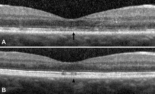
FIGURE 3 Humphrey visual field 30-2, right eye. Baseline visual field shows an enlarged blind spot and temporal depression (A). The visual field three months after treatment is improved (B).

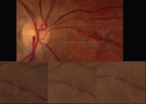
FIGURE 4 Fundus photographs, right eye. One week after initial presentation, a peri-papillary nematode is identified (top), and its movement through the subretinal space is observed (bottom).