Figures & data
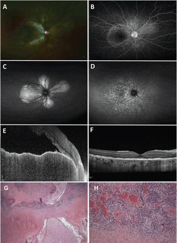
FIGURE 1 A 39-year-old male presented with blurred vision and loss of accommodation in the right eye 1 month after suffering a ruptured globe from facial trauma in the left eye. (A, B) Right eye. Dilated fundus examination showed mildly hyperemic optic nerve associated with exudative retinal detachment in the posterior pole (A) as well as multiple pinpoint areas of leakage on wide-field fluorescein angiography (B). (C, D) Right eye. Wide-field fundus autofluorescence revealed a petaloid pattern of hyperautofluorescence centered on the optic nerve corresponding to areas of exudative retinal detachment (C) at initial presentation that would later evolve to speckled areas of hyper- and hypoautofluorescence resembling leopard spots (D) following resolution. (E, F) Right eye. Enhanced depth spectral domain optical coherence tomography demonstrated a massively thickened choroid measuring more than 500 μm, irregularity of the retinal pigment epithelium, submacular fluid accumulation, disruption of the IS-OS line, and hyperreflective spots within the area of subretinal fluid accumulation (E). Three months following treatment, there was restoration of the normal foveal contour with some residual focal areas of photoreceptor loss (F). (G) Left eye. There is diffuse choroidal thickening with sheets of lymphocytes focally interrupted by nests of epithelioid histiocytes (1.25×, hematoxylin & eosin). No Dalén-Fuchs nodules were identified. (H) Left eye. Monomorphic lymphocytic infiltration of the choroid with occasional nests of epithelioid histiocytes (20×, hematoxylin & eosin). Note that the lymphocytic infiltrate involves the choriocapillaris.