869
Views
15
CrossRef citations to date
0
Altmetric
Letters to the Editor

Nested PCR-positive Tubercular Ampiginous Choroiditis: A Case Report

&
Pages 303-305 | Received 12 Sep 2011, Accepted 12 Apr 2012, Published online: 23 May 2012

Figures & data

FIGURE 1  (a) First follow-up fundus image of right eye showing whitish yellow choroiditis, new lesions, and atrophic lesions along vessels. (b) Late-phase FFA image of right eye showing the fuzzy margins due to leakage from the active lesions.
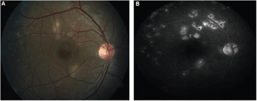
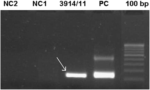
FIGURE 1  (a) First follow-up fundus image of right eye showing whitish yellow choroiditis, new lesions, and atrophic lesions along vessels. (b) Late-phase FFA image of right eye showing the fuzzy margins due to leakage from the active lesions.
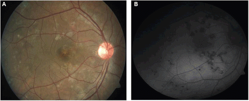
FIGURE 2  Gel photograph of positive nested PCR on aqueous sample for M.tb done against a positive (PC) and negative (NC) control.

FIGURE 2  Gel photograph of positive nested PCR on aqueous sample for M.tb done against a positive (PC) and negative (NC) control.

FIGURE 3  (a) Healed choroiditis lesions. (b) Fundus autofluorescence image shows no hyperautofluorescence within the lesions, suggesting no activity.

FIGURE 3  (a) Healed choroiditis lesions. (b) Fundus autofluorescence image shows no hyperautofluorescence within the lesions, suggesting no activity.

Reprints and Corporate Permissions

Please note: Selecting permissions does not provide access to the full text of the article, please see our help page How do I view content?

To request a reprint or corporate permissions for this article, please click on the relevant link below:

Academic Permissions

Please note: Selecting permissions does not provide access to the full text of the article, please see our help page How do I view content?

Obtain permissions instantly via Rightslink by clicking on the button below:

If you are unable to obtain permissions via Rightslink, please complete and submit this Permissions form. For more information, please visit our Permissions help page.