Figures & data
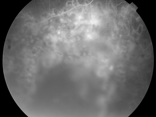
FIGURE 1 Fundus fluorescein angiogram of patient 1 following photocoagulation.
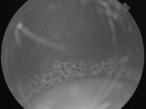
FIGURE 2 Fundus picture of patient 2 following photocoagulation. Note that laser spots form a barrier around the bullous retinoschisis.
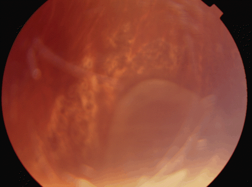
FIGURE 3 Fundus fluorescein angiogram of patient 3 following photocoagulation.