Figures & data
Figure 1. Fundus of left eye during vitrectomy. During vitreous surgery, a yellowish white retinal exudative lesion involving the posterior pole and whitening inside vascular arcades are observed, both of which are atypical findings of PDR. Proliferative membrane was around the disc.
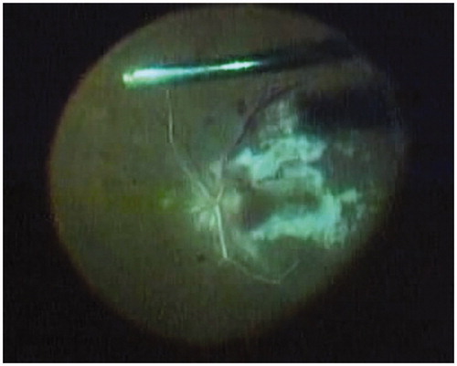
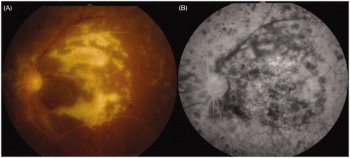
Figure 2. Fundus of left eye after treatment. (A) Macular color photograph and (B) macular fluorescein angiography. Ganciclovir therapy (600 mg/day) for 3 weeks achieved successful remission of the retinal lesion, but visual acuity of the left eye did not improve since macular atrophy had already progressed.