Figures & data
FIGURE 1. (a) Anterior segment photograph of left eye at presentation showing the presence of Koeppe nodules at the pupillary border, broad-based posterior synechiae, and hypopyon. (b) Anterior segment photograph showing resolution of hypopyon.
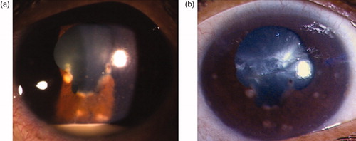
FIGURE 2. Histopathology section from cervical lymph node showing acid-fast bacilli (arrows).
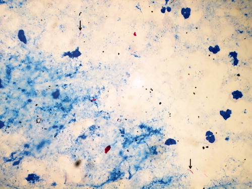
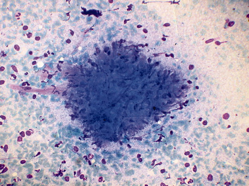
FIGURE 3. Cervical node biopsy showing granuloma with multinucleate giant cells, degenerated inflammatory cells, and necrosis.