Figures & data
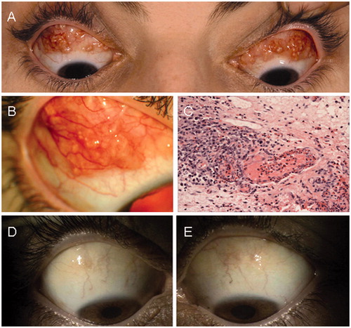
Figure 1. (A) The patient at presentation with bilateral multinodular involvement of the bulbar conjunctiva. (B) The appearance of the nodules on closer examination. (C) Conjunctival stroma with mixed inflammatory infiltrate and numerous eosinophils within a vessel wall (×20 objective, hematoxylin & eosin stain). (D, E) Resolution of the multinodular masses of the conjunctiva on the right and left, respectively.