Figures & data
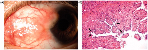
FIGURE 1. (A) Clinical picture shows solitary, lobulated conjunctival tumoral mass associated with conjunctival hyperemia encroaches on the cornea nasally. (B) Histopatology demonstrates suprabasal acantholitic separation (arrows) in the conjunctival epithelial cells and subepithelial congested vessels giving a papillary configuration to mucosa (hematoxylin–eosin stain × 200).