Figures & data
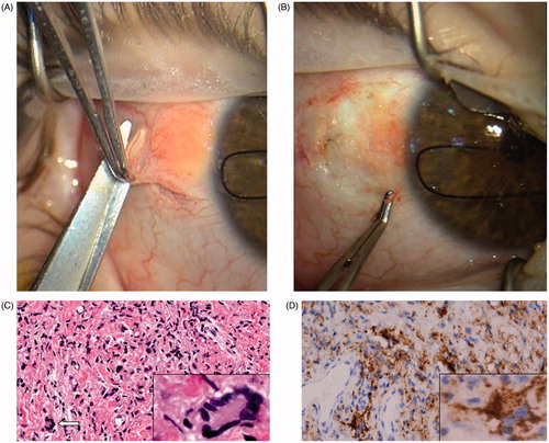
Figure 1. (A) Surgical excision of a subconjunctival yellow-orange lesion at the bulbar conjunctiva. (B) The subconjunctival mass has been dissected. The infiltration of the sclera was seen during the operation. (C) Microscopically, the specimen showed confluent histiocytes that were not particularly filled with lipid, scattered Touton giant cells (arrow and inset), and lymphocytes (hematoxylin–eosin, original magnification, ×100; inset, original magnification, ×400). (D) Diffuse CD68 positivity (macrophage marker) of the lesion (original magnification, ×100; inset magnification, ×400).