Figures & data
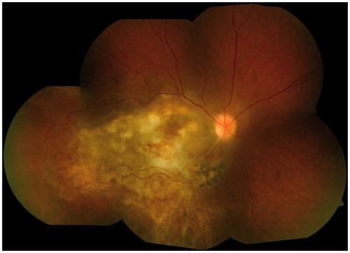
Figure 1. Fundus photograph (a) of right eye of the patient (case 1) at initial visit with visual acuity 6/60. There was significant vitritis with a yellowish placoid lesion in posterior pole and retinal hemorrhages in periphery.
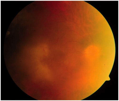
Figure 2. Subretinal aspirate (case 1) showing structures consistent with Toxoplasma gondii cysts with numerous bradyzoites, and a pigment-laden macrophage (May-Grumwald Giemsa, ×1000).
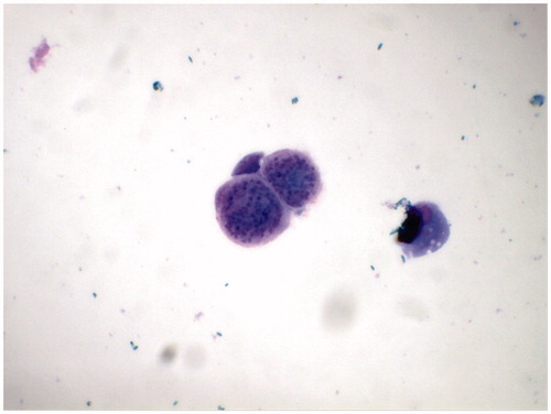
Figure 3. Fundus photograph (case 1) 3 days after surgery (pars plana vitrectomy + partial retinectomy + endolaser photocoagulation + silicon oil tamponade) showing attached retina in a silicon-oil filled eye.
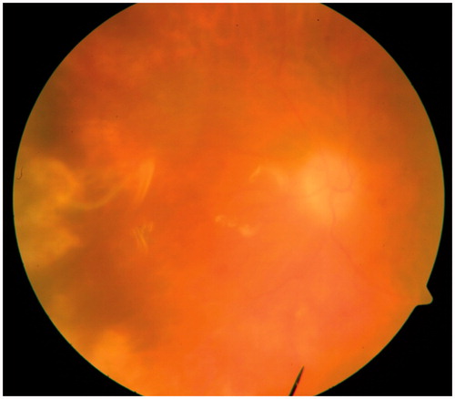
Figure 4. Fundus photograph of right eye (case 2) shows a widespread necrotizing retinitis with a healed choroiditis scar along the lower temporal arcade.
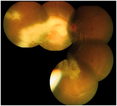
Figure 5. Vitreous smear (case 2) showing structures consistent with Toxoplasma gondii cysts in the main picture and in the inset (May-Grumwald Giemsa, ×1000).
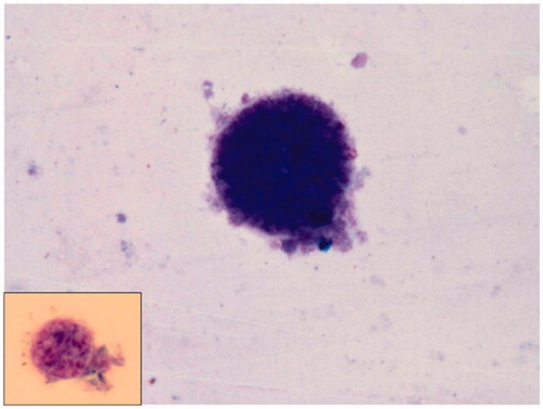
Figure 6. Fundus photograph (case 2) at 1 month follow-up showing healing lesions after anti-toxoplasma therapy was initiated.