Figures & data
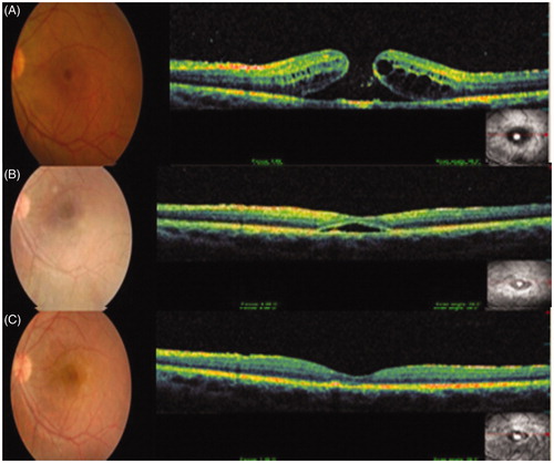
Figure 1. Color fundus photograph and spectral domain–optical coherence tomography (OPKO/OTI Spectral OCT/SLO, OPKO Health, Inc. Miami, Florida) images of the left eye show (A) a full-thickness macular hole before, (B) macular hole closure with subretinal fluid accumulation 1 month after, and (C) complete macular hole closure 4 months after the initiation of interferon alfa-2a therapy.