
Figures & data
Figure 1. Fundus photographs of both eyes on admission, showing vitreous opacity and serous retinal detachment in right eye, and hyperemic optic disk and serous retinal detachment in left eye.
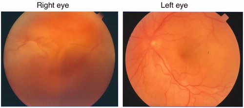
Figure 2. Denervation supersensivity test, after thirty minutes of one drop 0.125% pilocarpine, showed left pupil constriction from 6.7 to 4.1 mm.
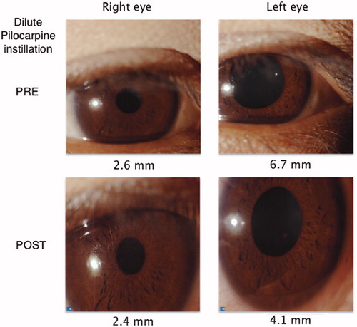
Table 1. Ocular manifestations.
Table 2. Systemic manifestations.