Figures & data
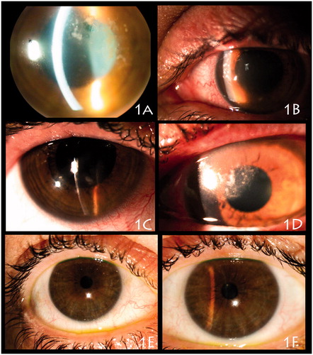
Figure 1. (A) Left eye episode diagnosed and treated as archipelago keratitis in 2007, with multiple microabscesses in the supero temporal cornea. (B) December 2009, left eye showing ulcerative blepharitis with superior lid oedema, erythema and erythematous macular lesions on the superior nasal lid skin; conjunctival hyperemia; multiple anterior stromal microabscesses situated on the superior nasal cornea and one infiltrate positioned in the inferior corneal; stromal diffuse infiltration and oedema. (C) November 2012, right eye showed mild conjunctival hyperemia, and 3 anterior stromal infiltrates associated with mild anterior diffuse stromal infiltration. (D) November 2012, her left eye showed superior lid oedema and an erythematous superior nasal lid lesion, meibomitis, and conjunctival injection. The left cornea presented with multiple stromal infiltrates positioned in the supero nasal cornea moving away from the limbus with a convexity border. (E–F) June 2013, right and left eyes showing eyelid margin without inflammation and cornea without scarring or corneal neovascularization (barely perceptible paracentral scarring, on both eyes, detected in 2009, are not seen in the pictures).